Each time somebody sneezes, and you see that aerosol drifting towards you, you know there is something in there that could potentially get you very sick. When you visit the doctor with that fever and runny nose, you are diagnosed with a viral infection, and hearing that, you know you are up for a lot of misery for the next few days! So, what are these viruses and how do they manage to cause so much chaos once they enter your body?
It all started with a bunch of really sick tobacco plants
In the late 1880s, two scientists – Adolf Mayer and Dmitri Ivanowsky, were working on a disease that had at that time become an epidemic – the Tobacco Mosaic Disease, which caused patchy, mosaic like discoloration of tobacco plant leaves. Remember, this was a time when advanced microscopy techniques were not yet developed to visualise the infectious agent. So, to study the causative agent, they crushed the infected leaves and obtained the fluid or sap and used this to infect other plants, with the understanding that this sap would contain whatever it is that caused the disease.
As expected, the sap did infect other plants. They then passed the sap through a filter that was known to filter out the smallest known infectious agent known at the time – bacteria. However, after filtration, the sap could still infect other plants! This was intriguing for the scientists and they concluded that the sap contained, perhaps some kind of soluble enzyme or toxin that could transmit the infection (Artenstein, 2012).
Not a toxin, but a contagium
It was only in 1895 that these conclusions were questioned, by a scientist named Martius Beijerinck, who conducted a slightly different set of experiments. He injected the filtered sap into different parts of the tobacco plant and noticed that the sap infected only the growing bits of the plant and not the parts that were fully grown.
Next, he allowed the filtered sap from the diseased plant (which contained the infectious agent) to sit for over three months, in the hopes that the nutrition in the sap should be able to propel to some extent, the growth of whatever was in there, and then used this sap to infect the plants. To his surprise he observed that the infection potential of the sap had neither increased nor lost. He then concluded that the infectious agent was actually a really small, soluble “contagium”, that he called ‘virus’. This virus, he said, was dependent on a cellular make up (like that of the tobacco plant) to infect and make more of itself, and could not divide outside it. Further he also noted that these viruses infected only growing, dividing cells and not tissues that were fully formed.
And then we could finally see them – the era of electron microscopy
It had become quite clear from these experiments that viruses wer too small to be captured by an ordinary light microscope. However, in the early 1900s, with the invention of the electron microscope, a whole new dimension was now visible (Goldsmith and Miller, 2009).
Researchers from all across the globe, working on different kinds of viruses started to examine them under the electron microscope and produced high quality images of these tiny specks (Goldsmith and Miller, 2009). What was evident, is that all viruses had one basic structure – an outer protein coat which was termed the capsid, inside which lay the genetic material (DNA or RNA), that coded for these proteins. Some viruses also had an envelope covering the protein coating, which they derived from the cell they were infecting3. These viruses could range anywhere between 20 nm to 300 nm in size, which means that they were so small that you could fit 10s to 100s of these into bacterial or human cells.
The mysterious life of a virus
However, there was something starkly mysterious about of these viruses. Yes, they did have a genetic material and a protein coating that protected this genetic material, but they completely lacked the protein and molecular machinery required to reproduce. Which means not only could they not replicate their own genetic material, but they could also not make their own protein coatings. They were completely disabled, unless they hopped on to something that actually had this machinery and stole it – like other living cells. On the other hand, they could be crystallised and stored in a nutritious medium for years on end, with little or no loss of virulence. The disability of these entities to do the most fundamental life process on their own and their ability to be resurrected back to life after long periods of crystallisation has made the scientific community debate for years if we can call a virus a ‘living’ entity at all. Although the debate is still ongoing, these entities can indeed be considered existing at the edge of life (Forterre, 2010).
Viruses have been known to infect every single cellular being on this planet4. Which means you and I are not alone when it comes to getting sick. Bacteria, protists like amoeba and fungi can get sick too!
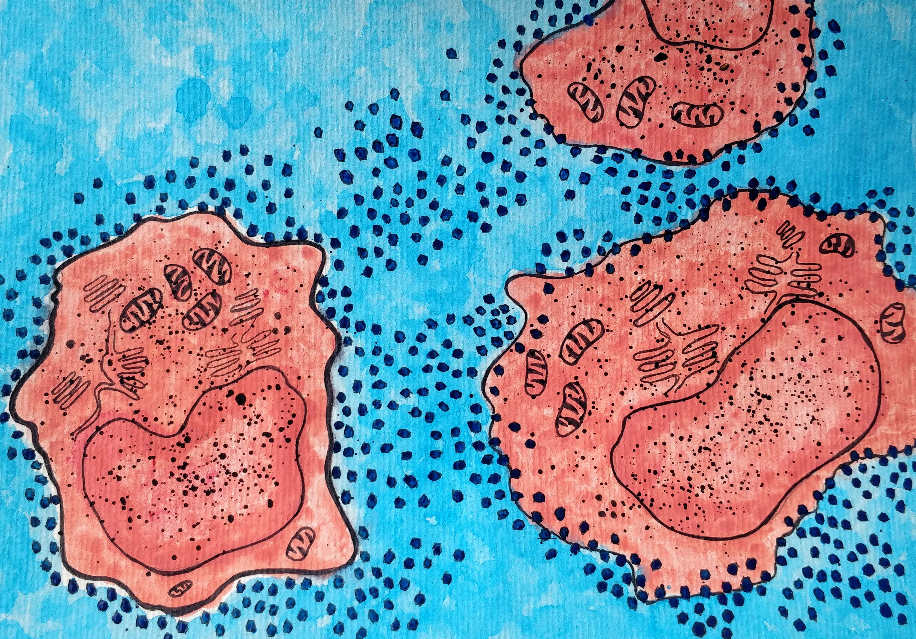
So how do viruses infect cells? All viruses follow one basic strategy. They first hop on and attach to the cellular membrane of the cell they are infecting. This is made possible by their outer proteins (the ones in the capsid or envelope), which recognise specific proteins on the surface of the cell membrane, thereby making specific viruses target only specific types of cells. That is the reason why, let’s say a plant virus, cannot infect an animal cell.
Next, they make their entry into the cell and release their genome (DNA/RNA) into the cell. Once in the cell they can access the cellular machinery which involves certain proteins and complex cellular organelles like ribosomes, which can now help make more copies of the viral genetic material and also help decode the information in its genome to make its proteins (like the ones that makes the viral capsid). The virus can better access this cellular machinery when the cell is dividing and hence the ability of most viruses to infect dividing cells (remember how the Tobacco Mosaic virus could only infect the dividing bits of the plant body?).
Finally, once numerous copies of the genetic material and the viral capsid proteins are made, the viral particles assemble – protein units gather to form the capsid, enclosing the genetic material. These particles are then released from the cell, and head out to infect more cells.
So, each time you sneeze, your mucous carries an army of these viral particles, just waiting to land into somebody else’s respiratory tract. Once it reaches there, it once again hijacks the cells there and carries forward its lineage!
Our weapons against viruses
The human population for centuries has been plagued by several viral infections, some reasonably harmless like the common cold and some fatal like AIDS, caused by the HIV virus. Decades of studies on viral genomes, protein structures and viral replication mechanisms have helped scientists a great deal in understanding how viruses propagate themselves and thereby helped develop strategies to combat infections. Antiviral drugs have been developed against many viruses such as those that cause Herpes, Influenza, Hepatitis and HIV, amongst many others, and this has helped reasonably contain the infection. The basic underlying mechanism of action of all these drugs is to stop the virus at any one of these stages:
Entry of the virus into the cell
Uncoating of the viral capsid once inside
Replication of viral genetic material (Billiau, 2007)
Assembly of the virus in the cell
Release of the viral particles form the cell
Another very efficient way of combating viruses would be through vaccinations. A vaccination is essentially a way of training your immune cells to face a prospective infection. In this case a small dose of the viral protein is injected into the body, which activates the immune cells of the body and gives it enough time to generates an appropriate immune response, without actually causing an infection. In this way if the actual virus were to ever infect the body, the immune cells would immediately recognise it and generate the right immune response.
We humans have come a long way, from studying the infectious filterable sap from tobacco leaves, to now being able to accurately, not just visualise those little specks but also understand how they work and inhibit their action. But what is most remarkable from these studies is that it has made us question the very nature of life. We as humans love categorising things – good and bad; heaven and hell; living and dead. But how do we categorise something that is neither here nor there? That is neither alive nor dead? Something that exists at the edge of life?
References
A. Artenstein, The discovery of viruses: advancing science and medicine by challenging dogma. International Journal of Infectious Diseases. 16, e470-e473 (2012). 10.1016/j.ijid.2012.03.005. context
C. Goldsmith and S. Miller, Modern Uses of Electron Microscopy for Detection of Viruses. Clinical Microbiology Reviews. 22, 552-563 (2009). 10.1128/cmr.00027-09. context
C. Goldsmith and S. Miller, Modern Uses of Electron Microscopy for Detection of Viruses. Clinical Microbiology Reviews. 22, 552-563 (2009). 10.1128/cmr.00027-09. context
P. Forterre, Defining Life: The Virus Viewpoint. Origins of Life and Evolution of Biospheres. 40, 151-160 (2010). 10.1007/s11084-010-9194-1. context