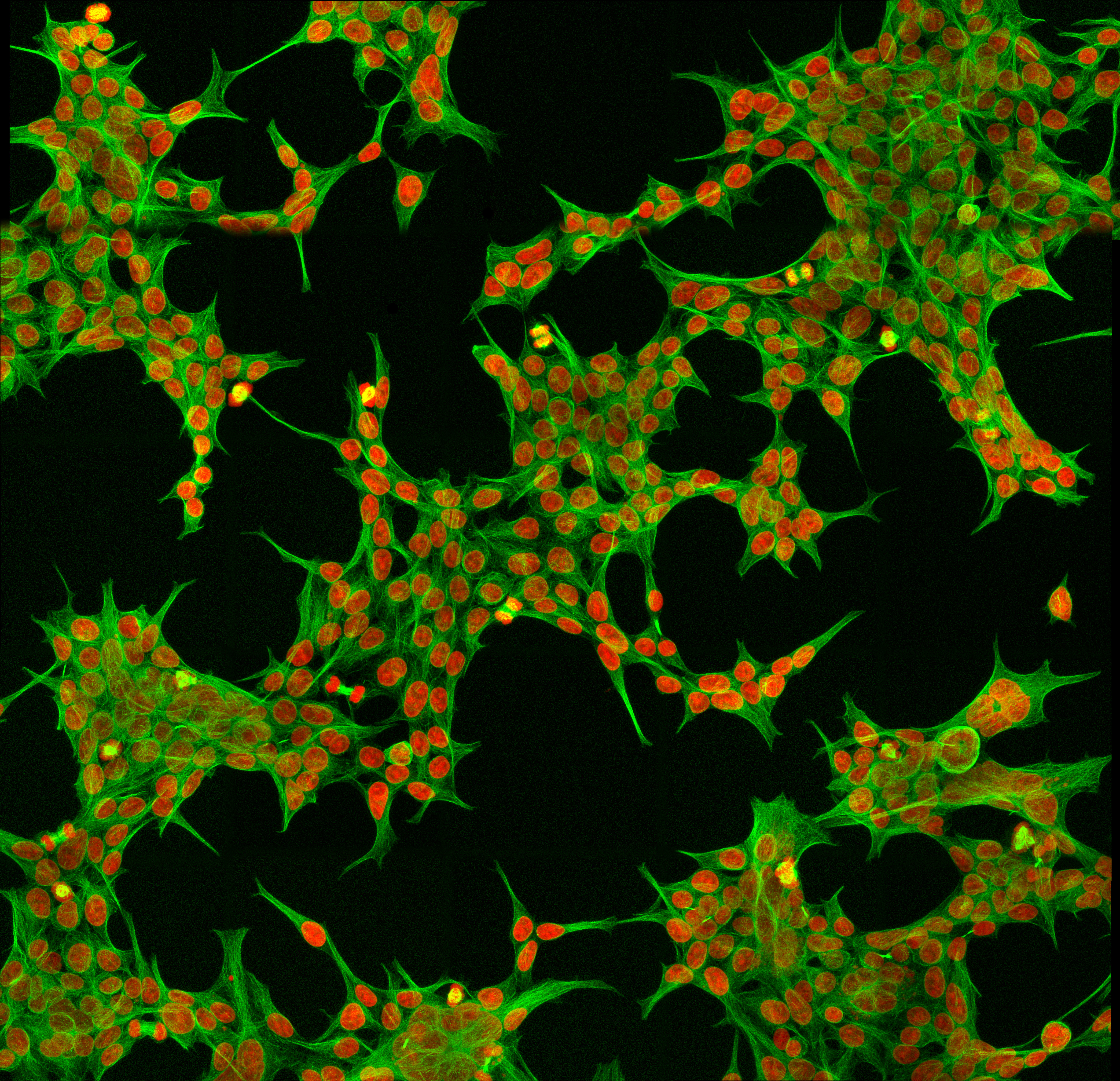
Human embryonic kidney cells as seen under the microscope. These cells have a typical shape which helps perform a specific function. Credit: Wikimedia.
If we carefully observed nature, we would see that we are surrounded by unique shapes. Take the beehive for example. You might have noticed that all the cells in a beehive are hexagonal in shape. This shape actually helps tightly pack the cells within the beehive and maximise the space for storage of honey.
Let’s take the fish as another example. Most fishes have a cylindrical body with tapering ends. This shape helps reduce friction as a fish swims through water.
What do both these examples have in common? - The shape helps perform a specific function.
Similarly, the cells in our body also have very unique shapes. Although textbooks often represent cells to be a circle or a rectangle, in reality our cells have some very different, interesting shapes. Just like the examples we saw before, these shapes too help the cells perform specific functions.
So let’s take a look at some of the uniquely shaped cells in our body and the functions they help perform.
Nerve cells (Neurons)
Neurons from a rat brain tissue as observed under a microscope. Each green and blue bulb-like structure is a neuron’s cell body. The red thread-like structures emerging from the cell body are its branches. These help in making connections with other neurons. Credit: Wikimedia.
The job of nerve cells or neurons is to transmit messages from different parts of the body to the spinal cord or brain. They also help transmit responses from the brain and spinal cord back to different body parts. These messages are in the form of electrical signals which are carried from one neuron to another, similar to electricity carried by electrical cables.
A typical neuron has a cell body with all its important organelles. Emerging from the cell body are a lot of short, and a few long branches. This kind of structure helps carry and distribute signals quickly, very often over large distances (Hagemann et al., 2022)
Red blood cells (RBCs)
Microscopy image of red blood cells. Credit: Ruihu Wang and Bin Fang (CC BY 3.0).
RBCs carry oxygen through our blood vessels and deliver it to different organs. An RBC is a round disc shaped cell with a dent in the center. This shape helps the cell to be flexible and allows it to twist or deform repeatedly as it makes its way through the narrow, twisted blood vessels (Diez-Silva et al., 2011).
Sickle shaped RBCs among normal RBCs found in sickle cell anemia. Credit: Wikimedia.
We see the importance of the RBC’s shape in the genetic disease, sickle cell anemia. The disease affects the shape of RBCs, making them sickle shaped, resembling a crescent moon. Sickle shaped RBCs are not as flexible as normal RBCs, making it much harder for them to move through the vessels. This can lead to a severe illness, causing extreme pain and even organ failure.
Skin cells
Tightly packed layers of cells in the skin. The outer layer is called the epidermis, and the inner layer is called the dermis. Credit: Wikimedia.
Our skin is made of sheets of cells which are stacked in many layers. This helps form a protective barrier between the outside world and the inside of our body.
Tetrakaidecahedron - A 14 sided geometric shape, with 6 rectangular and 8 hexagonal sides. This shape is proposed to be most effective in fitting cells tightly together. Credit: Wikimedia.
When scientists observed these cells under a powerful microscope, they found that they were tightly packed with the help of special connections. These connections were mainly possible because of the cells’ unique shape. The cells were observed to have a 14 sided geometric shape, with 6 rectangular sides and 8 hexagonal sides. This shape is called a tetrakaidecahedron and is proposed to be most effective in fitting cells tightly together (Yokouchi et al., 2016).
Neutrophils
A neutrophil chasing a bacterial cell while constantly changing its shape.
Neutrophils are a type of white blood cells (WBCs) which help ‘eat’ up invading pathogens in our body. Cells infected by pathogens release certain chemicals which attract neutrophils towards them. These chemicals help neutrophils locate where the pathogen is, so that they can move in that direction and engulf the pathogen.
Hence neutrophils are actually shape shifters. On sensing a pathogen, they form a front and back end, crawling towards the pathogen through the intricate gaps in tissues.
Sperm cells
Sperm cells of a bull as seen under a microscope. They show the typical head and tail structure. Credit: Wikimedia.
Sperms have evolved to be highly motile cells. After all, fertilising the egg is a challenging job, with millions of sperms competing with each other for the prize - the egg! Therefore, the quicker a sperm is, the better.
Sperms are hence extremely compact cells, with a head and a tail, somewhat resembling a tadpole. The head contains the DNA and no additional organelles. The tail contains a motile structure called the flagellum. This helps the sperm quickly propel itself towards the egg.
Conclusion
In all these examples what we see is that the cell’s shape is optimised for a specific function. But how do cells maintain their shape so strictly? This is a topic of interest for many scientists as it would help them better understand diseases where cells dramatically change their shape, like cancer.
References
C. Hagemann et al., Axonal Length Determines Distinct Homeostatic Phenotypes in Human iPSC Derived Motor Neurons on a Bioengineered Platform. Advanced Healthcare Materials. 11, (2022). 10.1002/adhm.202101817. context
M. Diez-Silva et al., Shape and Biomechanical Characteristics of Human Red Blood Cells in Health and Disease. MRS Bulletin. 35, 382-388 (2011). 10.1557/mrs2010.571. context
M. Yokouchi et al., Epidermal cell turnover across tight junctions based on Kelvin's tetrakaidecahedron cell shape. eLife. 5, (2016). 10.7554/elife.19593. context
.](/static/faf727467fc2f5237ee290b9cf867d60/b17f8/neurons.jpg "Neurons from a rat brain tissue as observed under a microscope. Each green and blue bulb-like structure is a neuron’s cell body. The red thread-like structures emerging from the cell body are its branches. These help in making connections with other neurons. Credit: [Wikimedia](https://commons.wikimedia.org/wiki/File:Chk-UCH1-GFAP-20X-1.jpg).")
.](/static/5e5ac594626b7a2424afa00445db7a47/db910/rbc-sem.png "Microscopy image of red blood cells. Credit: Ruihu Wang and Bin Fang [(CC BY 3.0)](https://creativecommons.org/licenses/by/3.0/).")
.](/static/f47d9ea0b92f0354566a8489a2e971ad/d4b53/sickle-cell-smear.jpg "Sickle shaped RBCs among normal RBCs found in sickle cell anemia. Credit: [Wikimedia](https://commons.wikimedia.org/wiki/File:Sickle-cell_smear_2015-09-10.jpg).")
.](/static/d73aa30928aa91e6ceaddd1365ecc383/a2510/epidermis-delimited.jpg "Tightly packed layers of cells in the skin. The outer layer is called the epidermis, and the inner layer is called the dermis. Credit: [Wikimedia](https://commons.wikimedia.org/wiki/File:Epidermis-delimited.JPG).")
.](/static/2452a186e2138b569f55645615a97881/81c8e/tetrakaidecahedron-6squares-8hexagons.png "Tetrakaidecahedron - A 14 sided geometric shape, with 6 rectangular and 8 hexagonal sides. This shape is proposed to be most effective in fitting cells tightly together. Credit: [Wikimedia](https://commons.wikimedia.org/wiki/File:Tetrakaidecahedron-6squares-8hexagons.png).")
.jpg).](/static/ef6d24115db8b461a59a5bc6698bcf8e/a12aa/sperm-cells-bull.jpg "Sperm cells of a bull as seen under a microscope. They show the typical head and tail structure. Credit: [Wikimedia](https://commons.wikimedia.org/wiki/File:Sperm_Cells_of_Bull_(430_times_magnification).jpg).")


.jpg)