A brief history of how our understanding of the cell/plasma membrane has evolved.
Table of Contents
- What is the Plasma Membrane?
- Groter and Grendel, 1925
- The Danielli – Davson Model, 1935
- The unit membrane model
- The fluid mosaic model of Singer and Nicolson, 1972
- References
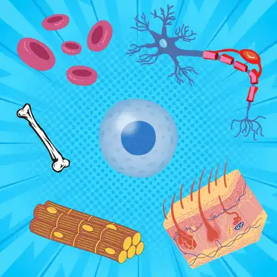
What is the Plasma Membrane?
The plasma membrane or cell membrane is a thin (around 75Å thick) membrane covering every cell in the human body. This membrane protects the cell, which is around 100 micrometer in diameter.
We know from a series of experiments conducted in the past few decades that this membrane is essentially composed of a phospholipid bilayer with proteins embedded within it (Robertson, 2004).
Let’s do a quick thought experiment! Imagine you were in the early 1900s; What basic experiments would you conduct to decipher the structure, functions and composition of this thin membrane, given that some basic information on the membrane was available to you:
- The membrane must have lipids in it, as lipid soluble factors easily enter the cells.
- Lipid molecules were amphiphilic with polar heads and non-polar tails.
Now that this exercise has gotten you wondering about different experimental assays, let’s look at how Groter and Grendel figured certain aspects of the cell membrane way back in 1925.
Groter and Grendel, 1925
Membranes have a lipid bilayer
In order to study the cell membrane, Groter and Grendel extracted RBCs from different animals, and isolated the lipids from the cells (Gorter and Grendel, 2004). They further calculated the surface area of these cells using the magnifying power of the microscope and some basic mathematical calculations.
They then suspended the extracted lipid molecules into a monolayer by dissolving the lipids in benzene, and added a few drops of the solution into water. Now they calculated the surface area occupied by this layer, and established that this area was twice the surface area of the cells under study. They hence concluded that the cell membrane must have a bilayer of lipid moieties, with their polar heads facing outwards (either towards the outside or towards the inside of the cell) and their non-polar fatty acid tails facing each other.
The theory however, had its pitfalls. For example:
- If the membrane was purely made of lipids, how did molecules insoluble in lipids get into the cell?
- Studies showed that the surface tension of the membrane was much lower than that of oil droplets, indicating that there must be some molecule associated with the membrane that lowers the surface tension.
Studies later proved this molecule to be a protein.
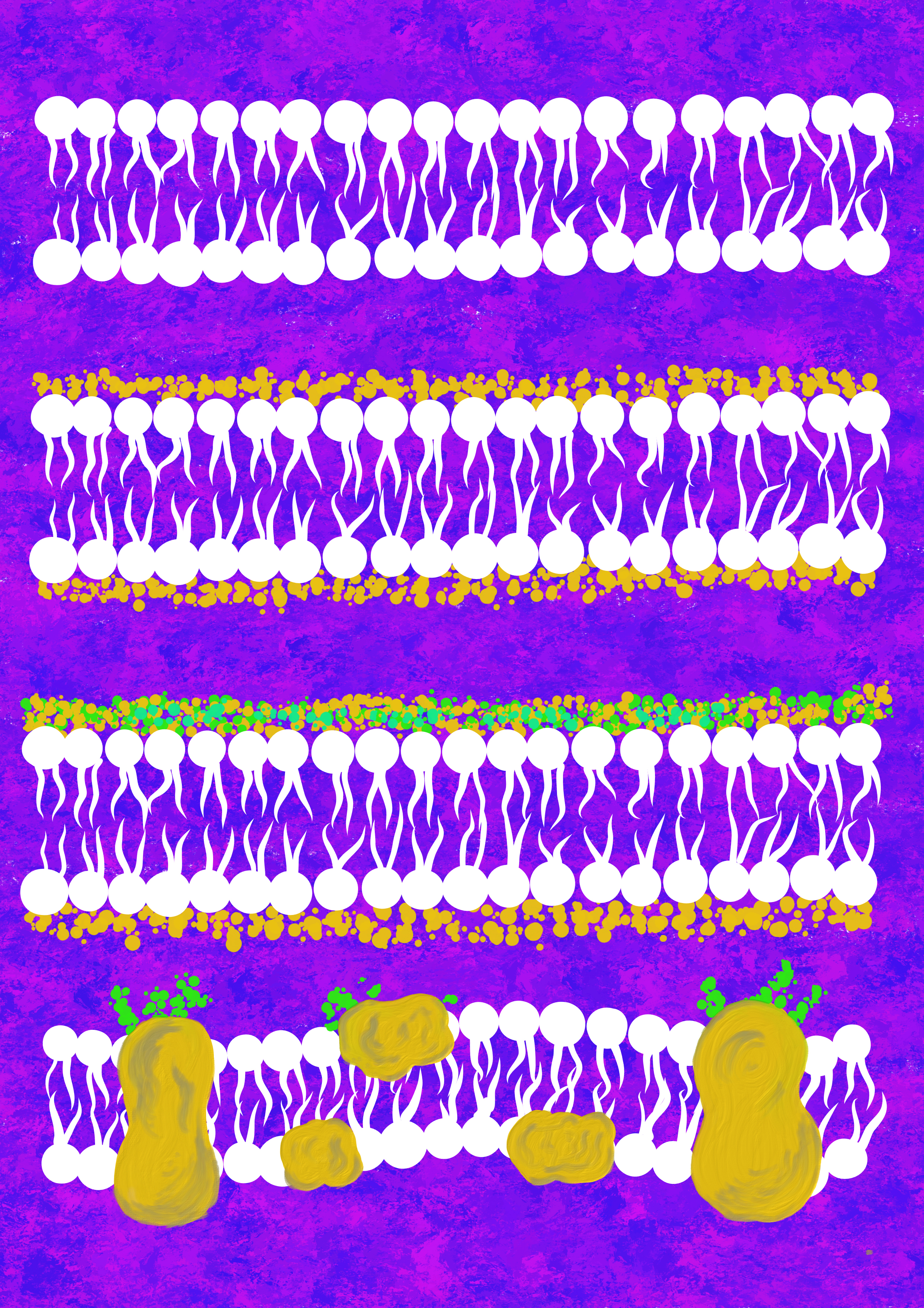
The Danielli – Davson Model, 1935
Not just lipids, membranes have proteins too
To clarify the existing criticism, Danielli and Davson proposed that cells were covered by a thin film of lipoidal substance (Danielli and Davson, 2005). By lipoidal, they meant a substance that was more soluble in hydrocarbons than in water. They suggested that the lipoidal substance had a lipoidal core bordered by monolayers of lipids, and adsorbed onto these, were protein monolayers.
Essentially the added element in this theory was the presence of proteins in the membrane, which tried to address the surface tension issue. This theory received prominence when the first electron microscope images of the cell membrane were produced in the 1950s. It showed the membrane to be a triple layered structure, made up of 2 dense regions (presumed to be made up of the hydrophilic heads of lipids and the protein coating) surrounding a light central region (presumed to be made up of the hydrophobic tails of the lipids).
The unit membrane model
Not just lipids and proteins, membranes have carbohydrates as well
Research rapidly boomed in this area due to the now available electron microscope. Series of experiments were conducted on a spectrum of cells, and all these experiments suggested one important thing – that all biological membranes were made of a similar bilayer, called the unit membrane. In other words, it suggested the universality of the lipid bilayer.
However, the unit membrane model also suggested one more very important thing – asymmetry. Electron microscopy images of cell membranes had shown a slight difference in the staining pattern between the outer and the inner surfaces. Biochemical and staining experiments proved that the outer layer had carbohydrates associated with them. Hence the unit membrane model also suggested that the cell membrane was asymmetrical, consisting of two dissimilar layers (One with carbohydrates and one without).
You might be beginning to think that we are coming down to the perfect model here…but not yet! This model too had to face its share of criticism. Fluorescence based experiments began to show that the proteins in the membrane did not essentially occupy the same position all the time – they moved! Not just this, freeze fracture etch electron microscopy – a method where in a frozen sample was fractured to study the internal structures, revealed that there existed considerably large sized particles ranging in size 50 Å – 100 Å within the cell membrane. So, proteins were not just present on the outside, they transversed the membrane too! (da Silva and Branton, 2004)
The fluid mosaic model of Singer and Nicolson, 1972
The cell membrane has fluidity
Singer and Nicolson finally gave the finishing touches to the long evolving cell membrane model (Singer and Nicolson, 2006). They suggested that the cell membrane had fluidity, and the components could move, much like a fluid mosaic. They also proposed that the membrane was not really bordered by protein molecules as suggested by previous models, but instead contained protein moieties dispersed in the membrane, just like icebergs floating in the water. They proposed that proteins were both peripheral as well as integral, with some spanning the membrane - transmembrane proteins. Some proteins on the outer surface had carbohydrate moieties linked to them.
This model holds to a great extent even today. Of course, the string of research that ensued after 1972 has revealed a plethora of details. An example for this would be studies that show that membranes are not as fluid as proposed previously. In fact, some proteins in the membrane are tethered to cellular cytoskeleton on the inside, and the extracellular matrix on the outside, making them immobile.
The cell membrane as we know now is not just a structure that separates the inside of the cell from the outside, but indeed partakes in important functions such as transport, attachment and signalling (Frye and Edidin, 2021). This makes it one of the most important components of the cell.
It is indeed intriguing to look back and see how over the past 100 years, our understanding of the cell membrane has transformed step by step, largely owing to the diligence and patience of the scientists, and the supporting technological advancements. It shows that it takes decades and sometimes centuries to paint the complete picture, and we can never actually be sure if the picture is indeed ‘complete’! Perhaps, that’s the beauty of science.
References
- J. Robertson, Membrane structure.. The Journal of cell biology. 91, 189s-204s (2004). 10.1083/jcb.91.3.189s. context
- E. Gorter and F. Grendel, ON BIMOLECULAR LAYERS OF LIPOIDS ON THE CHROMOCYTES OF THE BLOOD. Journal of Experimental Medicine. 41, 439-443 (2004). 10.1084/jem.41.4.439. context
- J. Danielli and H. Davson, A contribution to the theory of permeability of thin films. Journal of Cellular and Comparative Physiology. 5, 495-508 (2005). 10.1002/jcp.1030050409. context
- P. da Silva and D. Branton, MEMBRANE SPLITTING IN FREEZE-ETCHING. The Journal of Cell Biology. 45, 598-605 (2004). 10.1083/jcb.45.3.598. context
- S. Singer and G. Nicolson, The Fluid Mosaic Model of the Structure of Cell Membranes. Science. 175, 720-731 (2006). 10.1126/science.175.4023.720. context
- L. Frye and M. Edidin, The rapid intermixing of cell surface antigens after formation of mouse human heterokaryons. Journal of Cell Science. 7, 319-335 (2021). 10.1242/jcs.7.2.319. context