Researchers identified patterns of brain maturation in worms using electron microscopy, providing insights into brain development in humans.
Aug 05, 2021 · 2 min read
Table of Contents
- Research Question
- The Study
- Key Findings
- References
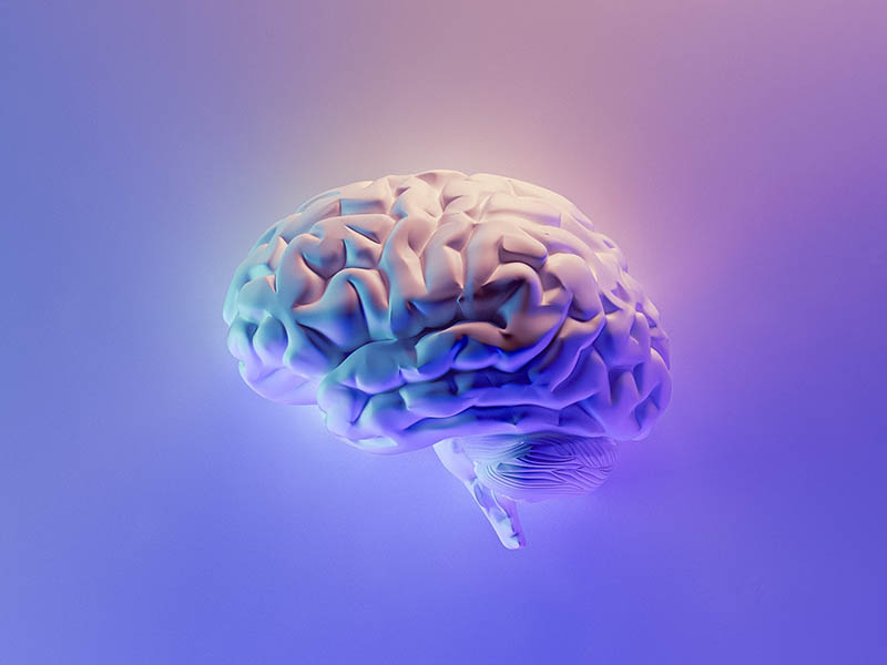
The human brain contains some 80-90 billion neurons or nerve cells (Azevedo et al., 2009). These neurons are the functional units of the brain. Their function depends on creating, maintaining and strengthening connections between other neurons. Connections between neurons are facilitated by special structures in the neurons called synapses.
A synapse permits the passage of electrical/chemical signals to the neighbouring neuron thereby establishing a connection. A complex network of such synapses and connections constitutes the brain. How this network matures during brain development was not understood.
Now, researchers from Canada and the USA have shed some light on brain development from birth to adulthood. They used a model worm and electron microscopy to reveal the details. The results of their research were published in the journal Nature (Witvliet et al., 2021). In this report, FROMTBOT summarises their key findings for you.
Research Question
What are the principles that govern the development of the brain?
The Study
The researchers used Caenorhabditis elegans (a tiny worm) as a model system to study neuron connections during brain development. They used a special form of electron microscopy where serial, ultra-thin sections of the worm’s brain were imaged. This allowed them to reconstruct the whole brain in a detailed fashion. The reconstruction was performed in 8 genetically identical worms. They then measured the number of synapses and connections between neurons in the brains at various stages of development.
Key Findings
- As the body develops, the brain increases in size while maintaining overall shape and geometry. The length of neurons increased proportionally to body length.
- The brain develops by creating new synapses and connections. New synapses made already existing connections stronger. Neurons that were already in contact at birth showed higher probability to form a connection during maturation.
- Newer synapses and connections during development segregate groups of neurons into functional modules or sub-networks (for example sensory, head movement, body movement etc). The number of modules increases with age. This indicates increase in complexity of the brain.
- Neuron connections across individuals can be classified as:
- Stable: maintained from birth to adulthood
- Dynamic: connections existed but their number and strength varied across individuals
- Variable: connections were found only in some individuals. The set of all variable connections in an individual were unique to that individual.
- The generation of new synapses preferentially creates connections from sensory to motor. “Thus, one global pattern of brain maturation augments signal flow from sensation to action, making the brain more reflexive with age”, the authors write in the paper.
The lead author of the study Dr. Mei Zhen said in a press release, “This is the first time that an entire brain’s structure is deduced and compared across developmental stages, from birth to adulthood.”
“These new findings have powerful implications for the fundamental rules that allow the brain’s developmental maturation to take place,” she added.
References
- F. Azevedo et al., Equal numbers of neuronal and nonneuronal cells make the human brain an isometrically scaled‐up primate brain. Journal of Comparative Neurology. 513, 532-541 (2009). 10.1002/cne.21974. context
- D. Witvliet et al., Connectomes across development reveal principles of brain maturation. Nature. 596, 257-261 (2021). 10.1038/s41586-021-03778-8. context